Virtual Scattering Lab
Matthias B. Hullin,
Martin Fuchs,
Ivo Ihrke,
Hans-Peter Seidel,
Hendrik P. A. Lensch
MPI Informatik
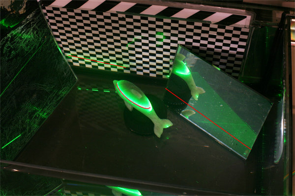
Test scene for light sheet range scanning in scattering media.
From left to right: opaque checkerboard surface, translucent stone dolphin,
mirror behind glass. The intersection line between light sheet and surfaces is marked in red.
Single vs. multiple scattering
In our SIGGRAPH 2008 paper "Fluorescent Immersion Range Scanning", we introduce a method to scan the 3D geometry of objects by making light rays visible as they propagate through a surrounding volume. Apparently, we are not the first ones who had this idea, see e.g. the ICCV 2005 presentation to the project "Structured Light in Scattering Media" by Narasimhan et al. (slide #12 in the Powerpoint file). On this web page, we compare the performance of dilute milk and a fluorescent dye with regard to light sheet range scanning. It turns out that finding a suitable concentration of milk is somewhere between very hard and impossible (which would explain why the idea has not been published so far). A fluorescent dye, however, immediately produces clear and sharp results regardless of the concentration.
Move the slider to change the milk concentration.
Move the slider to change the amount of fluorescent dye.
Results
Comparison of dilute milk and Eosin Y with regard to their scattering properties gives the following results:
- The color of the scattered light is changed in the fluorescent medium, whereas it remains the same (=laser wavelength) in milk.
- The image quality depends strongly on the concentration of milk, while it is nearly independent of the Eosin concentration. For higher concentrations of milk, multiple scattering reduces the contrast significantly, while in the fluorescent solution there is virtually no multiple scattering at all.
- In the case of dilute milk, the "brightness gap" of the light sheet at the mirror surface can only be observed for very thin solutions. However, in the presence of bright reflections from the object surfaces (e.g. the dolphin) this is hardly detectable at all. Using Eosin, the longpass filter removes all direct reflections and transmits only the scattered light.
Explanation
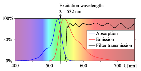
Fluorescence is an "inelastic" scattering process where incoming photons are absorbed by dye molecules and emitted at a larger wavelength. This process can only be repeated if the emitted photon has enough energy (wavelength short enough) to be absorbed again. Given the small overlap between absorption and emission spectra (see diagram), it is highly unlikely that a photon undergoes a fluorescent scattering event multiple times. Since we use an excitation wavelength close to the long-wavelength tail of the absorption spectrum, the majority of photons observed by the camera has been scattered in the medium exactly once.
Back to the project page
 Fluorescent Immersion Range Scanning
Bibliography
Matthias B. Hullin, Martin Fuchs, Ivo Ihrke, Hans-Peter Seidel, Hendrik P. A. Lensch, "Fluorescent Immersion Range Scanning" , ACM Trans. on Graphics (Proceedings of ACM SIGGRAPH 2008), Volume 27, Issue 3 (August 2008).